Clinical Evidence Behind the First FDA Cleared Al-Enabled Device for Skin Cancer Assessment

Validated across clinical studies demonstrating non-invasive assessment of suspicious skin lesions, with real-time quantitative cancer risk result. 

Clinical Evidence & Research

Review the clinical studies behind DermaSensor, including study objectives, trial design, enrollment data, key findings, and conclusions.

Study 1

DERM-SUCCESS FDA Pivotal Study: Primary Care Physician Use of Elastic Scattering Spectroscopy on Skin Lesions Suggestive of Skin Cancer

This study investigated the sensitivity and specificity of a noninvasive, hand-held DermaSensor in evaluating skin lesions compared to the in-person clinical evaluation by primary care physicians (PCPs).

  • DermaSensor demonstrated high sensitivity of 95.5% in detecting all skin cancer types when compared to the gold standard of histopathologic examination, with similar sensitivity and specificity across all Fitzpatrick skin type subgroups.
  • NPV of the device was 96.6%, meaning a negative “monitor” device result had only a 3.4% chance of being a false negative, i.e. any of the three cancer types.
  • The overall positive predictive value (PPV) for an “investigate further” result was 16.6% (NNB of 6:1).
  • The device sensitivity and overall accuracy (i.e. AUC) were both found to be superior to those of the PCPs.
  • By pairing high sensitivity with clinical exam findings, the device may rule out 20.7% of lesions from needing further evaluation, reducing patient anxiety and optimizing healthcare resource allocation.
  • Likelihood of malignancy increased significantly with increasing spectral scores. For scores of 1-3, PPV was 6%, which increased to 18% for scores of 4-7 and 40% for scores of 8-10.

This blinded, prospective, multi-center study was conducted at 22 primary care study sites across the United States (18 sites) and Australia (4 sites). Patients with lesions suggestive of skin cancer were clinically assessed by PCPs and then evaluated by the DermaSensor. Patients and PCPs were blinded to device output. All lesions enrolled were biopsied per physician assessments and standard of care. Each lesion’s diagnosis involved 2-5 dermatopathologists, dependent on pathology and discordance. Statistical analyses after study unblinding included standard diagnostic test parameters of the device for detecting skin cancer as well as the influence of lesion and patient factors on device performance.

During study enrollment, five lesions (0.3%) were excluded due to device data capture issues and five lesions (0.3%) due to lack of dermatopathology consensus. A total of 1,005 patients with 1,579 lesions suggestive of skin cancer were enrolled. Among the patients enrolled, 51.4% were female with a mean age of 59 years, and 72.5% of patients were Fitzpatrick Skin Type I-III.

The DermaSensor device is an easy-to-use, point-of-care, hand-held skin cancer adjunctive diagnostic device with high sensitivity and NPV to help inform PCP decision-making about skin lesions suspicious for cancer that need further evaluation and those that may be monitored. Coupled with clinical exam findings, this device may aid PCPs to improve clinical decisions about suspicious skin lesions (i.e., to refer, or monitor), with this study suggesting device use could rule out 20.7% of suspicious lesions from needing further evaluation. With the device PPV reaching as high as 61%, the device can also help inform which lesions merit urgent evaluation by dermatologists.
Study 2

DERM-SUCCESS FDA Pivotal Study: A Multi-Reader Multi-Case Evaluation of Primary Care Physicians’ Skin Cancer Detection Using AI-Enabled Elastic Scattering Spectroscopy

The aim of this study was to assess and compare the diagnosis and management performance of primary care physicians (PCPs) with and without the use of the handheld DermaSensor in detecting skin cancer.

  • Improvement in Sensitivity: Management sensitivity increased significantly from 82.0% to 91.4% (p=0.003) with device output. Diagnostic sensitivity increased significantly from 71.1% to 81.7% with device output (p=0.008). Specificity decreased non-significantly to 60.9% to 54.7% (p=0.190) for diagnosis and 44.2% to 32.4% (p=0.026) for referrals.
  • 52% False Negative Reduction: Skin cancer referrals decreased from 18.0% to 8.6% with device use, a relative decrease of 52%.
  • Improvement in Physician Confidence: Physicians reporting high confidence in their management assessment increased from 36.8% to 53.4%.
  • Improvement in Area-Under-the-Curve: Overall management performance (i.e., AUC) increased from 0.708 to 0.762, and it increased from 0.567 to 0.682 for lesions which physicians reported low confidence in their unassisted management decision.

In this clinical utility study, 108 PCPs evaluated 50 skin lesions (25 malignant, 25 benign), with and without ESS device output. For each case, high-resolution digital clinical images, the patient’s clinical information, including prior skin cancer history, risk factors, and physical examination results were provided. The PCPs completed a questionnaire about their diagnosis of the lesion, their recommended management decision, and their confidence level in their management decision for each case. Sensitivity and specificity of PCP diagnostic and management with and without the device output were calculated.

PCP participants included U.S. board-certified internal and family medicine physicians with an even distribution of years in practice (range:1-21+ years). PCPs were recruited from across the U.S., including urban and rural areas.

Management sensitivity increased significantly from 82.0% to 91.4% (p=0.003) with device output. Thus, PCPs’ false negative referrals decreased from 18.0% to 8.6%, a relative decrease in false negatives of 52%. Diagnostic sensitivity increased significantly from 71.1% to 81.7% with device output (p=0.008). Specificity was found to decrease non-significantly from 60.9% to 54.7% (p=0.190) for diagnosis and 44.2% to 32.4% (p=0.026) for referrals. Additionally, the effectiveness analyses observed an increase in PCPs’ overall diagnostic performance (i.e. AUC) and confidence level in their management decisions with the use of the handheld DermaSensor. This technology improves sensitivity and inherent validity of skin cancer diagnosis and management. The use of the ESS device in a primary care setting is further supported by a reduction in the subjectivity of the PCPs regarding their evaluations and the limited training required for its use, as well as the increased diagnostic and management performance.

Study 3

DERM-ASSESS III FDA Pivotal Study: Validation of a Handheld Elastic-Scattering Spectroscopy Device on Lesions Suggestive of Melanoma

This study aimed to validate whether the use of an ESS point-of-care test can detect melanoma when dermatologists are evaluating lesions that were concerning for melanoma.

  • The 10 dermatology study centers biopsied all lesions that were suspicious of melanoma based on their standard of care clinical and dermoscopic assessment and decision making.
  • DermaSensor was able to identify 96% of melanomas when compared to dermatopathology consensus results (at least two dermatopathologists reviewed each high-risk melanocytic lesion case); for both melanomas and several atypical nevi, the device sensitivity was 91%.
  • Overall device specificity was 33%, thus DermaSensor could have effectively ruled out 33% of benign biopsied lesions as benign while detecting 96% of melanomas.
  • The overall negative predictive value (NPV) for a “monitor” result was 98% for melanoma, i.e. a negative result had less than a 2% chance of being melanoma.
  • The positive predictive value (PPV), i.e. the likelihood a lesion was melanoma for a positive “investigate further” result, was 10% for low 1-3 scores (NNB of 10:1), 21% for mid 4-7 scores (NNB of 5:1), and 47% for high 8-10 scores (NNB of 2.1:1).
  • The overall device accuracy (i.e. Area Under the Curve or AUC) of 0.76 was comparable to that of 0.75 for the Dermatologists.

Ten dermatology study centers, across the US and Australia, scanned lesions that they found concerning for melanoma. All dermatologists were blinded to the device results. Gold standard comparison for performance of the device and dermatologists was the biopsy result with multiple dermatopathologist review when consensus was not reached during the primary review process. High resolution digital images and the patient’s clinical information, including prior skin cancer history, risk factors and physical exam results, were recorded for each case. After clinical evaluation, dermatologists reported their diagnosis and confidence level, which provided the physician comparison data. The results evaluated were sensitivity, specificity, Negative Predictive Value for melanoma, melanoma + severely atypical nevi, and all high-risk lesions. Area Under the Curve (AUC) was also calculated and modeled and compared between the study dermatologists and the device performance.

A total of 311 patients with 440 biopsied lesions were evaluated by the study dermatologists, device and dermatopathology results.

The use of the handheld DermaSensor by physicians, in addition to clinical evaluation, may improve melanoma detection. The device was able to identify 96% of melanomas when compared to dermatopathology results.

While this study was conducted by melanoma specialists, given the device’s similar overall performance to these specialists and its simple, non-invasive use, there is potential for the device to be used to help rule in or out lesion referrals for primary care physicians.

Study 4

Clinical Capabilities of a Handheld Elastic Scattering Spectroscopy – Artificial Intelligence Device as an Adjunctive Tool for Evaluating Skin Cancer in Skin of Color

To evaluate DermaSensor’s performance in patients with skin of color, this sub-analysis of the DERM-SUCCESS study compared device sensitivity and specificity in patients of Fitzpatrick skin type I-III and IV-VI subgroups.

  • Unlike tools that use clinical and/or dermoscopic images, which many publications have shown are subject to skin type bias, skin color has not been found to impact DermaSensor results which has the potential to augment PCPs exams and improve skin cancer detection capabilities across skin types.
  • Overall sensitivity was similar between Fitzpatrick skin types I-III and IV -VI, with 96.5% (95% CI: 92.1-98.5%) and 92.2% (95% CI: 82.1-96.8%), respectively. Sensitivity was consistent across Fitzpatrick skin types.
  • The specificity of the DermaSensor for Fitzpatrick skin types I-III was 18.7% (95% CI: 16.2-21.5%) and 25.1% (95% CI 20.9-29.7%) for types IV-VI. The device has the potential to avoid 18.7% to 25.1% of unnecessary care that was provided for suspicious yet benign lesions.

Ten dermatology study centers, across the US and Australia, scanned lesions that they found concerning for melanoma. All dermatologists were blinded to the device results. Gold standard comparison for performance of the device and dermatologists was the biopsy result with multiple dermatopathologist review when consensus was not reached during the primary review process. High resolution digital images and the patient’s clinical information, including prior skin cancer history, risk factors and physical exam results, were recorded for each case. After clinical evaluation, dermatologists reported their diagnosis and confidence level, which provided the physician comparison data. The results evaluated were sensitivity, specificity, Negative Predictive Value for melanoma, melanoma + severely atypical nevi, and all high-risk lesions. Area Under the Curve (AUC) was also calculated and modeled and compared between the study dermatologists and the device performance.

A total of 311 patients with 440 biopsied lesions were evaluated by the study dermatologists, device and dermatopathology results.

The use of the handheld DermaSensor by physicians, in addition to clinical evaluation, may improve melanoma detection. The device was able to identify 96% of melanomas when compared to dermatopathology results.

While this study was conducted by melanoma specialists, given the device’s similar overall performance to these specialists and its simple, non-invasive use, there is potential for the device to be used to help rule in or out lesion referrals for primary care physicians.

Study 5

Enhancing Diagnostic Precision in Primary Care: A Multireader Multicase (MRMC) Study of an AI-Powered Handheld Elastic Scattering Spectroscopy Device for Informed Referral Decisions in Melanoma Evaluation

Given the importance of PCP decision making regarding lesions suspicious of skin cancer, particularly melanoma, it is postulated that use of the ESS device may assist PCPs in referral decision making and enhance provider confidence in managing these cases. Here, we describe the findings of a multireader multicase (MRMC) study to assess the referral performance of PCPs when evaluating lesions suggestive of melanoma with and without the aid of the ESS device.

  • The study met the primary endpoint, and a statistical test for superiority found that physicians device-aided accuracy (i.e. AUROC) was significantly higher than the unaided accuracy.
  • Aided PCP AUROC increased 4.1% for all skin cancers, achieving non-inferiority (p<0.001) and superiority (p=0.036)
  • Aided PCP sensitivity for all skin cancers increased 8.1%, achieving non-inferiority (p<0.001) and superiority (p<0.001)
  • Aided PCP sensitivity for melanoma increased 8.8%, achieving non-inferiority (p<0.001) and superiority (p<0.001)
  • Aided PCP specificity decreased by 5.6%, achieving its non-inferiority margin (p<0.001)

This melanoma-focused reader study was a web-based, MRMC investigation using clinical information, digital images, and ESS device result data. PCPs were asked to perform 200 reads for 100 skin lesion cases, each presented without and then with ESS device output.

The study examined two aspects of PCP decisions concerning skin evaluation using the device: (1) the impact of the device result on the PCPs’ management decision on whether the lesion should be referred for further evaluation by a dermatologist, and (2) the device’s impact on the PCP’s diagnostic assessment of whether the lesion was malignant or benign.

A total of 118 PCP readers, none of whom participated in prior studies pertaining to the device, completed the study and were eligible for the effectiveness analysis.

The study met the primary endpoint; the area under the receiver operating characteristic (AUROC) of the PCPs aided with the device was 0.671 compared with the AUROC unaided by the device of 0.630, a significant increase (p=0.036).

The device significantly improved physicians’ accuracy in managing lesions suggestive of melanoma, as well as significant increases in sensitivity for all skin cancers and melanoma, while not significantly raising the rate of unnecessary referrals. Moreover, the physicians reported increased confidence in their assessments with the availability of the device result. As such, the device can improve PCP decision making and confidence when managing
lesions suggestive of melanoma.

By enhancing PCP management accuracy and confidence, this FDA-approved device has the potential to optimize resource utilization by minimizing unnecessary referrals and biopsies, saving healthcare dollars, and reducing patient morbidity and concern, while helping to prioritize access for patients with high-risk lesions. Given the rising rates of skin cancer in the US and limited access to dermatologic care, this device has the potential to make a significant impact in skin cancer detection in the primary care setting.

Study 6

Prospective Evaluation of an AI-enabled Elastic Scattering Spectroscopy Device for Triage of Patient-identified Skin Lesions in Dermatology Clinics

This investigator-initiated, matched comparison study aimed to evaluate device performance on patient-identified lesions in a dermatology setting to assess its utility for screening lesions appropriate for referral across clinical contexts.

  • As the first independent investigator-initiated study conducted with DermaSensor in the US, this study at UPMC found that the overall diagnostic accuracy, i.e., Area Under the Curve (AUC), was 0.79, which was identical to the AUC in the FDA pivotal study with over 1,000 patients and 22 primary care study centers [8].
  • Sensitivity and specificity of the device were 100% and 9.4%, respectively, using Investigate Further vs Monitor as the cut point for positive vs negative. The publication also reported that sensitivity was 86.4% and specificity was 67.2% when considering 7-10 results as positive and 0-6 results as negative.
  • 59.5% of the Investigate Further false positive results were still considered high risk lesions in that they were actively managed or treated by the study dermatologists.

The authors prospectively evaluated 150 patients presenting with self-identified lesions of concern to 3 outpatient dermatology offices. Diagnostic accuracy of device readout (classifying lesions as either “Monitor,” alone, or “Investigate Further,” alongside a score of 1 to 10, indicating spectral similarity to malignant tissue) was compared to blinded dermatologist management decisions and histopathology.

The study included 150 lesions in 72 males and 78 females, with a mean age of 59.6 years.

Malignant lesions were identified in 14.7% of cases (22/150), with melanoma, BCC, and SCC accounting for 3, 9, and 10 cases, respectively.

This study suggests that the ESS device, as a complementary tool, may enhance triage and decision-making for patient-identified lesions to be employed by non-specialists in any clinical setting.

The ESS device identified 138 lesions as “Investigate Further” and 12 as “Monitor.” Sensitivity for detecting malignant lesions was 100%, negative predictive value (NPV) was 100%, specificity was 9.4%, and positive predictive value (PPV) was 15.9%. For lesions with a spectral score of 7 to 10, sensitivity was 86.4%, NPV 96.6%, with specificity increasing to 67.2% and PPV to 31.1%. Compared to dermatologists’ decisions, sensitivity was 95.5%, NPV 75%, specificity 10.7%, and PPV 45.7% (Table II)

  • These results were similar to those found in the DERM-SUCCESS pivotal study for
    sensitivity and AUROC as well as lesion distribution across spectral scores.
  • Applicability of the device in the PCP setting can be enhanced with development of clear management guidelines for specific lesion types.

Regulatory Milestones for DermaSensor

  • Only skin cancer device available to PCPs that provides any kind of automated risk assessment
  • Only automated skin cancer device indicated for use with all common skin cancers
  • Only currently available automated skin cancer device that uses Elastic Scattering Spectroscopy ESS
Learn More About Our Regulatory Journey

Podcasts

Listen and gain insights from experts on clinical perspectives, skin cancer detection, Al-assisted diagnostics, and patient care.

Show More

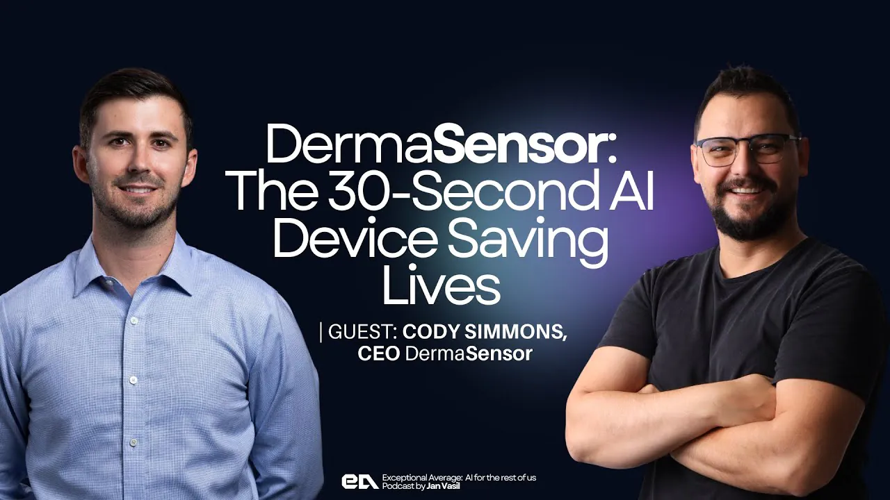
The Excepional Average Podcast: 30-Second Cancer Detection: The AI Device That’s Changing Medicine | Cody Simmons CEO ‪@Derma-Sensor‬

The Future of Skin Cancer Detection with Cody Simmons, CEO of DermaSensor

The Medical AI Podcast: Episode 48: Skin cancer: how can AI help with detection? With Cody Simmons, CEO of DermaSensor

Cody Simmons, CEO & Co-Founder of DermaSensor joins Dr. Felix Beacher.

Skin Cancer Detection with DermaSensor | Beyond The Blade Podcast (S2 – Episode 39)

Listen to Cody Simmons, CEO & Co-Founder of DermaSensor with Dr. Alexander Zuriarrain—renowned plastic surgeon, founder of Hydrology and Zuri Plastic Surgery in Miami, and an early adopter of DermaSensor—on the Beyond the Blade podcast.

80-0009.17