How DermaSensor Works with ESS and AI
Skin cancer identification often starts with a visual exam. But visual assessment alone can’t detect changes happening below the skin’s surface.
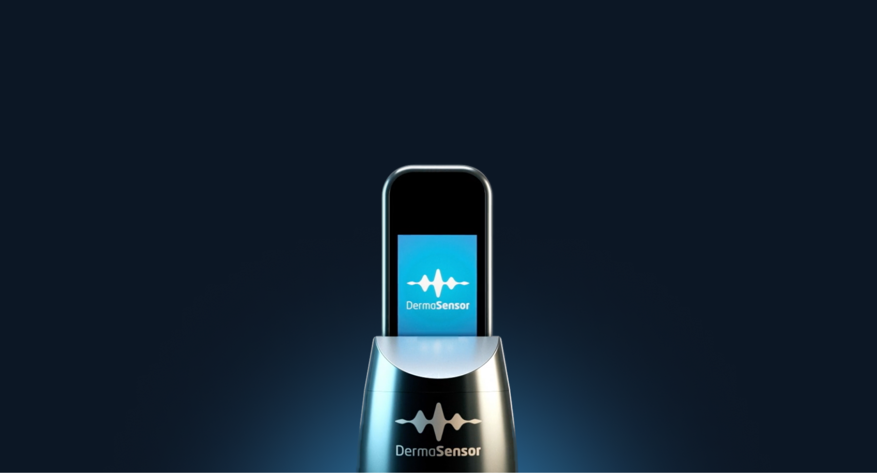
DermaSensor™uses Elastic Scattering Spectroscopy (ESS) combined with machine learning analysis to support skin cancer risk detection at the point of care. The technology is designed to provide objective data about suspicious lesions during a clinical visit, helping healthcare professionals assess risk beyond what may be visible to the naked eye.
ESS evaluates cellular and subcellular structures beneath the skin’s surface, identifying patterns associated with malignancy that may not be detectable through visual examination alone.
Learn how ESS skin cancer risk detection and AI analysis work at the point of care, and review the clinical evidence supporting this technology.
Quick Facts About ESS Skin Cancer Detection and AI:
ESS Technology
DermaSensor uses Elastic Scattering Spectroscopy (ESS) to analyze cellular and sub-cellular features.
Non-Invasive
Non-invasive, point-and-click spectral recordings.
AI Algorithm
AI algorithm that has been trained and validated by over 20,000 spectral scans.
Real-Time Output
Provides real-time output on a handheld device: "Investigate Further" or "Monitor".
For Clinical Settings
Designed for use by healthcare professionals in clinical settings.
Three Skin Cancers
Intended to aid in the evaluation of lesions suggestive of melanoma, basal cell carcinoma (BCC), and squamous cell carcinoma (SCC).
Not for Direct Diagnosis
Not intended for direct diagnosis of skin cancer.
*Clinical evidence upon request.
What Is ESS?
Elastic Scattering Spectroscopy (ESS) is a form of sub-diffuse reflectance spectroscopy that detects disease-associated changes at the cellular and subcellular level by analyzing how light interacts with tissue architecture. Unlike taking images of lesions, ESS translates tissue morphology directly into spectral features that correlate with histopathologic characteristics. ESS has been evaluated with 30+ peer-reviewed publications on clinical studies that show its ability to distinguish malignant from benign tissue across different tissue types.
Here’s how DermaSensor applies ESS at the point of care:
Light & Tissue Interaction
DermaSensor delivers short pulses (~30 microseconds) of broadband white light spanning 300-900 nm wavelengths. When this light penetrates tissue, photons scatter from refractive-index gradients associated with micro- and nano-scale structures, including nuclear size, chromatin condensation, cellular architecture, and collagen organization. An adjacent collection fiber captures backscattered photons and conveys them to a microspectrometer.
Spectral Analysis & AI Pattern Recognition
The system records a spectral signature of backscattered intensity versus wavelength. Malignant transformation alters cellular and subcellular structures, creating distinct optical signatures in the ESS spectrum. Lesions with different morphology, cellular, and subcellular features have different densities. The recorded spectrum undergoes preprocessing (smoothing and downsampling) before analysis by the proprietary neural network machine learning algorithm, which compares the pattern against training data from over 2,000 histopathologically confirmed lesions.
Clinical Use & Workflow Integration
Time-gated detection enables the device to function in ambient room light without requiring darkening, making it practical for clinical workflows. The entire measurement takes seconds and requires no tissue preparation.
*DermaSensor is intended for use by qualified healthcare professionals in clinical settings only. It is not intended for at-home or personal use.
Ready to See How DermaSensor Works in Practice?
How the DermaSensor Device Captures Spectral Data
The process begins with simple contact:
1. Device Contact
The handheld device is placed directly on the suspicious lesion.
2. Spectral Recording
Five (5) non-invasive spectral recordings are taken.
3. Light Scattering
The device measures how light scatters within the tissue.
4. AI Analysis
The spectral data is analyzed by an onboard AI algorithm.
The entire process is an immediate point-and-click device that is designed for use at the point of care. There is no tissue removal. No incision. No specimen collection.
The device records the spectral data of the lesion and compares it to patterns learned from thousands of known malignant and benign lesions.
How AI Is Used to Detect Skin Cancer Risk
Once captured, the spectral data is analyzed by a trained algorithm. The algorithm has been trained and validated using:
Over 20,000 spectral scans
More than 4,500 skin lesions
Histologically confirmed melanoma, BCC, and SCC
Benign lesions, including unbiopsied lesions diagnosed by board-certified dermatologists
The AI compares the lesion's spectral profile to known patterns.
It then generates one of two outputs:
Output 1
Investigate Further
Output 2
Monitor
For lesions classified as "Investigate Further," the device also provides a score from 1 to 10. This score reflects the degree of spectral similarity to malignant lesions in studies, with 10 representing the highest degree.
This output is designed to assist in determining whether a lesion may require further clinical care.
*DermaSensor is not intended to provide a direct diagnosis of skin cancer.
Real-Time Output at the Point of Care
After analysis, the results are displayed on the device immediately.
The output is designed to augment clinical decision-making by providing objective information during the patient visit.
In clinical studies:
- Overall device sensitivity for detecting all three common skin cancers was 96%
- In a primary care study, device sensitivity was 95.5% compared to 83.0% for participating PCPs
- The Negative Predictive Value (NPV) for a “Monitor” result was 96.6% in the primary care study
- In a dermatology study, diagnostic sensitivity was 96% for melanoma
- Sensitivity was 93% for all malignant lesions
Use of device output during clinical evaluation was associated with statistically significant improvements in both management sensitivity and diagnostic sensitivity.
Ready to See How DermaSensor Works in Your Practice?
*These findings are based on prospective, multi-center clinical studies. Clinical evidence is available upon request.
From Technology to Clinical Impact
Suspicious skin lesion assessments are made during a patient visit. Monitoring, biopsy, or referral must be determined in real time.
DermaSensor delivers structured output during that moment of decision-making, without disrupting workflow. No tissue removal. No added procedural steps.
Lesion Support
Supports assessment of lesions suspicious for melanoma, BCC, and SCC
Clinical Decisions
Provides information that can support clinical management and referral decisions during the visit
Clinical Evidence
Incorporating device output into evaluation can enhance management and diagnostic sensitivity
Important Safety Information
DermaSensor is:
- Intended to act as an adjunctive tool that uses ESS technology to support clinicians in evaluating lesions suspicious of melanoma or non-melanoma skin cancer (NMSC).
- Designed for use only by physicians and qualified healthcare professionals trained in the clinical assessment of skin lesions.
- Not intended for direct diagnosis of skin cancer.
For clinicians evaluating suspicious skin lesions, DermaSensor provides objective spectral data to support, not replace, clinical judgment.

How DermaSensor Works - FAQs
Frequently Asked Questions About How DermaSensor Works
How does DermaSensor work?
DermaSensor uses Elastic Scattering Spectroscopy (ESS) to analyze backscattered light from cellular and sub-cellular features of a suspicious lesion.
A locked, validated AI algorithm processes the data and delivers an immediate result of either “Investigate Further” or “Monitor”. The device is not intended for direct diagnosis and should be used alongside clinical assessment.
Who is DermaSensor designed for?
DermaSensor is intended for healthcare professionals who are not specialists. It is designed for use in clinical settings to support evaluation and management decisions.
The device is not intended for patient self-use.
How does DermaSensor support skin cancer risk detection?
DermaSensor analyzes spectral patterns to detect cellular changes associated with melanoma, BCC, and SCC, providing an immediate point-of-care result of either “Investigate Further” or “Monitor”. It is designed as an adjunctive tool used alongside clinical assessment, not as a replacement for clinical judgment. In FDA-submitted studies, PCP sensitivity increased from 80.5-82.0% unaided to 86.3-91.4% device-aided.
What results does the DermaSensor device provide?
DermaSensor provides one of two outputs:“Investigate Further” or “Monitor.”, For lesions classified as “Investigate Further,” a 1-10 score reflects the degree of similarity to malignant lesions observed in validation studies.
See How DermaSensor Works
Request a demo to explore how DermaSensor uses ESS technology to deliver objective, point-of-care results in seconds.