Background and Objective
Access to a dermatologist for diagnosis and management of skin cancer is challenging, as around 40% of the United States population live in areas that have limited access to a dermatologist [1]. This puts primary care physicians (PCPs) in an integral role for early detection of skin cancers, as early detection of these malignancies is imperative to decreasing morbidity, mortality, and cost to the patient [2].
Elastic Scattering Spectroscopy (ESS), an optical tissue sampling technique, distinguishes between normal and abnormal tissue in vivo without the need to remove tissue.

A handheld device that employs ESS and artificial intelligence was developed as an adjunctive tool for PCPs, to aid in their management of lesions suspicious for skin cancer. The technology has been shown to have skin cancer sensitivity over 90% in various prospective, multi-center studies when compared to gold standard dermatopathology results [3-5].
The aim of this study was to assess and compare the diagnosis and management performance of PCPs with and without the use of the handheld ESS device in detecting skin cancer.
Materials & Methods
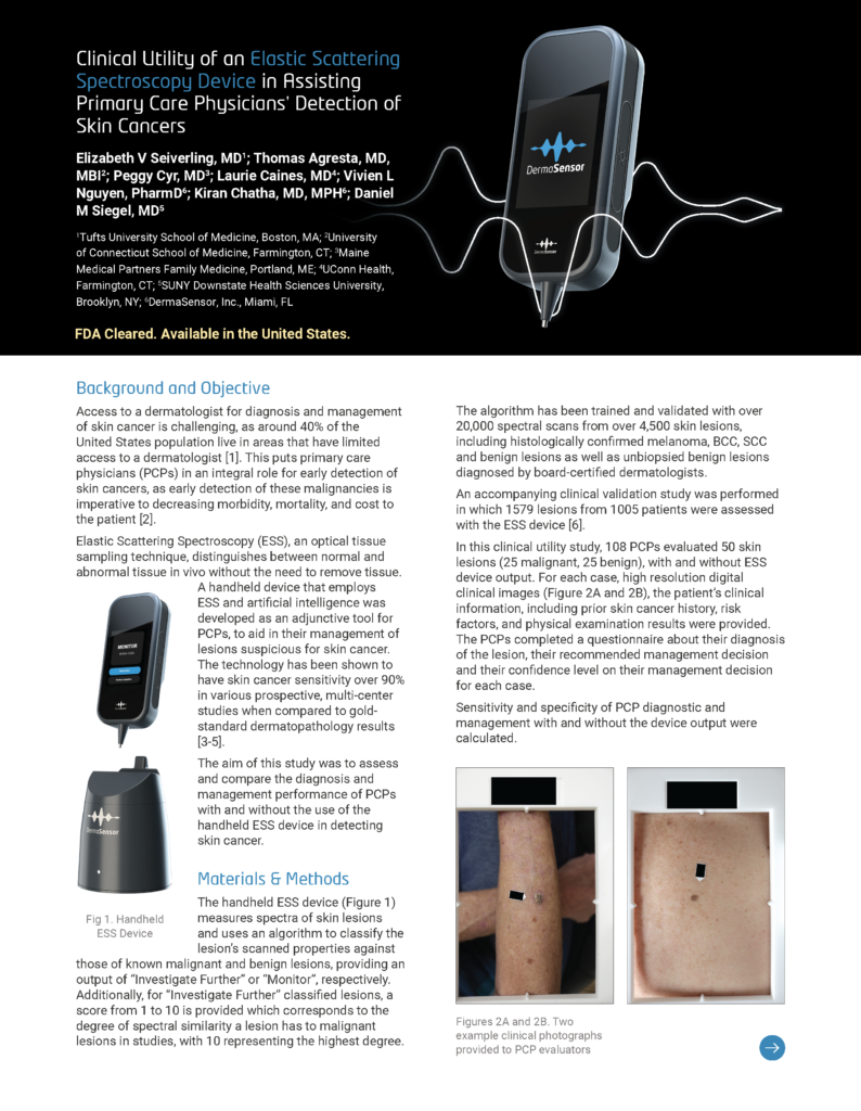
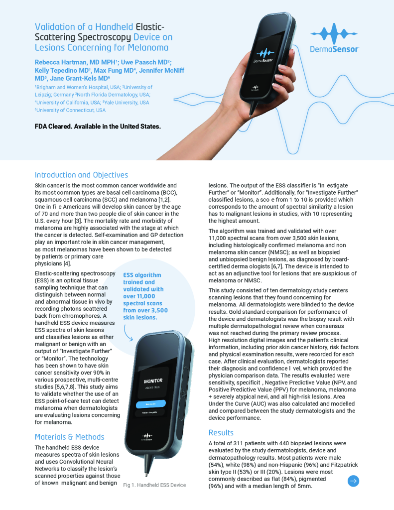
The handheld ESS device (Figure 1) measures spectra of skin lesions and uses an algorithm to classify the lesion’s scanned properties against those of known malignant and benign lesions, providing an output of “Investigate Further” or “Monitor”, respectively. Additionally, for “Investigate Further” classified lesions, a score from 1 to 10 is provided which corresponds to the degree of spectral similarity a lesion has to malignant lesions in studies, with 10 representing the highest degree.
The algorithm has been trained and validated with over 20,000 spectral scans from over 4,500 skin lesions, including histologically confirmed melanoma, BCC, SCC and benign lesions as well as unbiopsied benign lesions diagnosed by board-certified dermatologists.
An accompanying clinical validation study was performed in which 1579 lesions from 1005 patients were assessed with the ESS device [6].
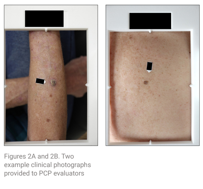
In this clinical utility study, 108 PCPs evaluated 50 skin lesions (25 malignant, 25 benign), with and without ESS device output. For each case, high resolution digital clinical images (Figure 2A and 2B), the patient’s clinical information, including prior skin cancer history, risk factors, and physical examination results were provided. The PCPs completed a questionnaire about their diagnosis of the lesion, their recommended management decision and their confidence level on their management decision for each case.
Sensitivity and specificity of PCP diagnostic and management with and without the device output were calculated.
PCP Participants
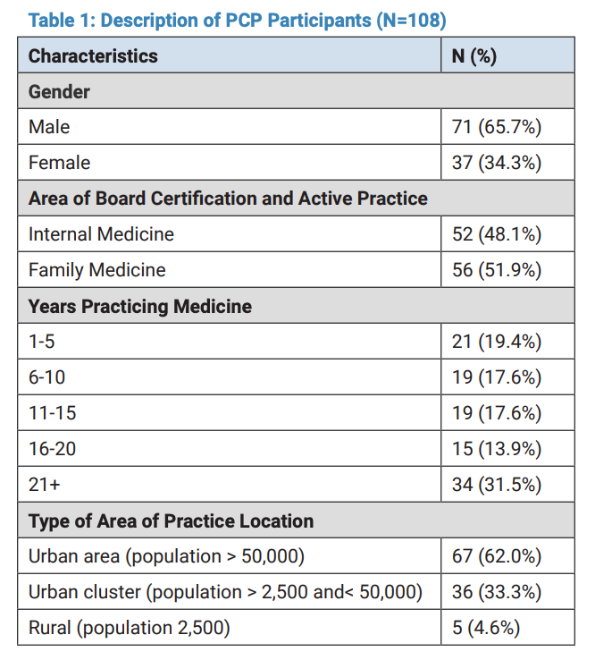
PCP participants (Table 1) included U.S. board-certified internal and family medicine physicians with an even distribution of years in practice (range: 1-21+ years). PCPs were recruited from across the U.S., including urban and rural areas.
Lesion Characteristics
The 50 lesions (Table 2) evaluated by PCPs were dispersed across the body (22.0% head, 26.0% upper and 10.0% lower extremities, 32.0% trunk) had a mixture of surfaces (66.0% elevated vs. 34.0% flat), textures (46.0% smooth vs. 54.0% rough), and pigmentation (48.0% light vs. 52% dark). Pathology of lesions were matched to the clinical study prevalence.
Results
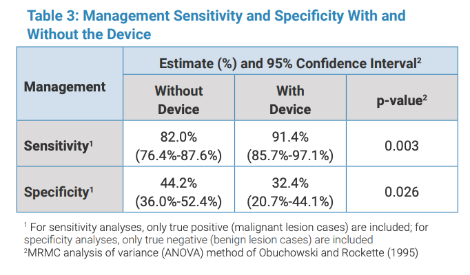
Management sensitivity increased significantly from 82.0% to 91.4% (p=0.003) with device output (Table 3). Diagnostic sensitivity increased significantly from 71.1% to 81.7% with device output (p=0.008) (Table 4).
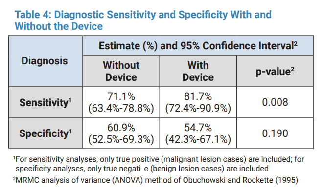
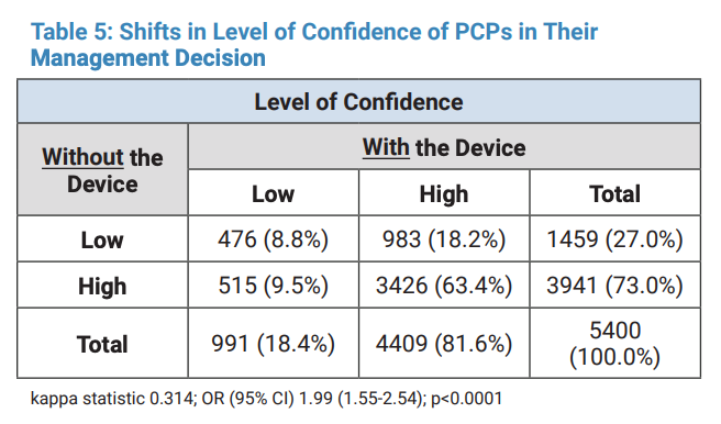
Specificities decreased from 60.9% to 54.7% (p=0.190) for diagnosis and 44.2% to 32.4% (p=0.026) for referrals. Overall diagnostic performance (i.e. AUC) increased from 0.685 to 0.727. Physicians reporting high confidence in their assessments increased from 73.0% to 81.6% (<0.0001) with device output (Table 5).
Conclusion
Use of the ESS device by PCPs significantly improved both diagnostic sensitivity (from 71% to 82%) and management sensitivity (from 82% to 91%). There were clinically acceptable decreases in associated specificities. Additionally, the effectiveness analyses observed an increase in PCPs’ overall diagnostic performance (i.e. AUC) and confidence level in their management decisions with the use of the handheld ESS device. The findings suggest the use of the ESS device output improves PCP skin cancer detection and confidence in skin lesion evaluation and management.
References
- Hester T, Thomas R, Cederna, J. et al. Increasing Access to Specialized Dermatology Care: A Retrospective Study Investigating Clinical Operation and Impact of a University-Affiliated ree Clinic. Dermatol Ther (Heidelb). 2021;11(1):105-115.)
- Perera E, Gnaneswaran N, Jennens R, Sinclair R. Malignant Melanoma. Healthcare (Basel). 2013;2(1):1- 19.
- Rodriguez-Diaz E, et al. Optical Spectroscopy as a Method for Skin Cancer Risk Assessment. Photochem Photobiol. 2019;95(6):1441-1445.
- Thames T et al. Clinical Utility of a Handheld Elastic Scattering Spectroscopy Tool and Machine Learning on the Diagnosis and Management of Skin Cancer. Poster Presentation, STFM Annual Spring Conference, April 30-May 4, 2022.
- Hartman R, Tepedino K, Fung, MA, McNiff, JM, Patrick G, Nguyen V, Chatha K, Grant-Kels J. Validation of a Handheld Elastic-scattering Spectroscopy Device on Lesions Suggestive of Melanoma. J Dermatol Physician Assist. 2022 Fall; 16(4):51.
- Merry SP, McCormick, B, Leffell D, Chatha K, Croghan I. Clinical Performance of Novel Elastic Scattering Spectroscopy (ESS) in Detection of Skin Cancer: A Blinded, Prospective, Multi-Center Clinical Trial [Initial Results]. Poster Presentation, Innovations in Dermatology, Las Vegas NV, Nov 3-6, 2022.
- Clinical Performance of Novel Elastic Scattering Spectroscopy (ESS) in Detection of Skin Cancer: A Blinded, Prospective, Multi-Center Clinical Trial [Initial Results]. Poster Presentation, Innovations in Dermatology, Las Vegas NV, Nov 3-6, 2022.
Disclosures
This study was supported by a grant from DermaSensor Inc. Drs. Nguyen and Chatha are employees of DermaSensor, Inc.
Adapted from Seiverling EV, Agresta T, Cyr P, Caines L, Nguyen VL, Chatha K, Siegel DM. Clinical Utility of an Elastic Scattering Spectroscopy Device in Assisting Primary Care Physician’s Detection of Skin Cancers. Poster Presentation, Maui Derm Hawaii Conference, Wailea, HI, January 24-28, 2023.